

Dynamic MRI of the Wrist for TFCC Evaluation Using Esaote O-scan MRI
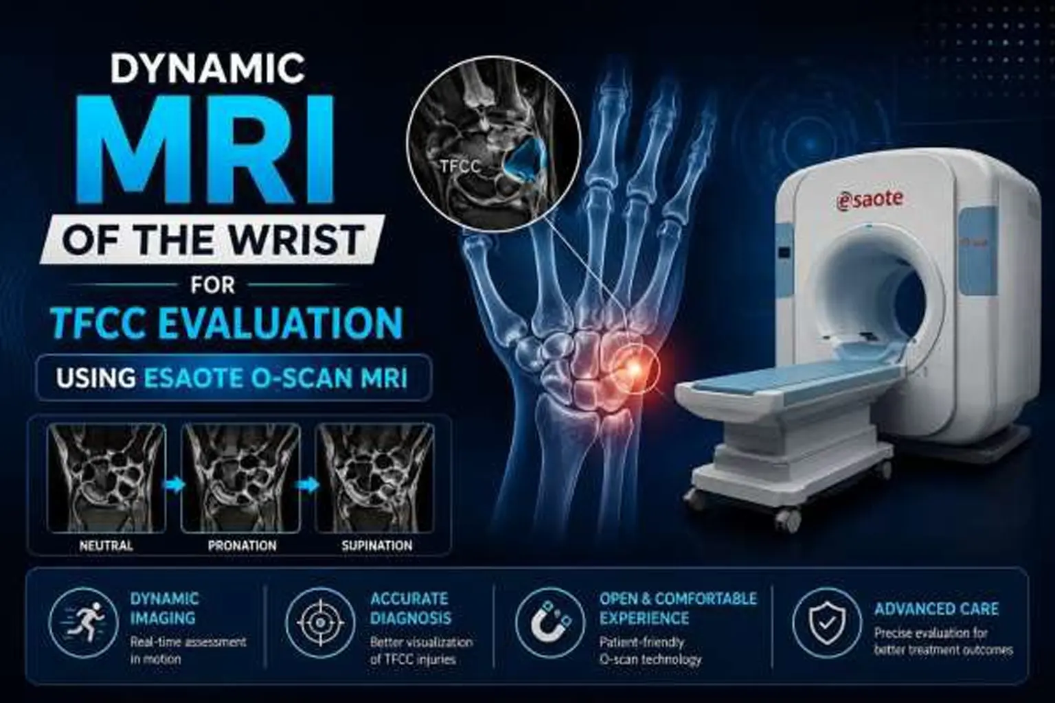
Wrist pain, clicking, weakness, and instability can significantly affect daily activities, sports performance, and work. One of the important structures associated with these symptoms is the Triangular Fibrocartilage Complex (TFCC).
While conventional MRI provides detailed anatomical information, some TFCC injuries and wrist instability may become more apparent when the wrist is examined under controlled stress. Dynamic MRI of the wrist offers an additional functional assessment by evaluating the wrist in specific loading positions.
At the Center for Sports Injury, under the clinical guidance of Dr. Pushpinder Singh Bajaj, Founder, advanced imaging approaches can help in the evaluation of sports-related and musculoskeletal wrist problems. The Esaote O-scan Dedicated Extremity MRI System
can be used for specialized dynamic MRI wrist stress studies, including horizontal stress view and upright vertical stress view, when clinically indicated.
What Is the TFCC?
The Triangular Fibrocartilage Complex (TFCC) is a group of cartilage and ligamentous structures located on the little-finger side of the wrist.
The TFCC helps:
- Stabilize the distal radioulnar joint (DRUJ)
- Support wrist movement
- Distribute forces across the wrist
- Maintain stability during gripping and rotation
- Absorb and transmit load through the wrist
TFCC injuries can occur following falls, twisting injuries, sports trauma, repetitive wrist loading, or degenerative changes.
Common symptoms may include ulnar-sided wrist pain, clicking, weakness, reduced grip strength, and a sensation of instability.
What Is Dynamic MRI of the Wrist?
A conventional MRI generally examines the wrist in a neutral or relatively relaxed position. This provides excellent anatomical information but may not demonstrate every functional abnormality.
Dynamic MRI evaluates the wrist in selected positions under controlled stress or loading conditions. This can provide additional information about how structures such as the TFCC and DRUJ behave when the wrist is subjected to functional forces.
For patients whose symptoms suggest instability but whose routine imaging findings are subtle or inconclusive, dynamic assessment may provide useful additional information.
Two Dynamic Stress Methods for Wrist and TFCC Evaluation
The Dynamic Wrist MRI protocol can include two specialized stress positions.
1. Horizontal Stress View
During the Horizontal Stress View, the wrist is positioned horizontally, and controlled stress is applied according to the imaging protocol.
This view can help assess:
- Peripheral TFCC abnormalities
- Central TFCC defects
- Distal Radioulnar Joint (DRUJ) alignment
- Changes in joint spacing under stress
- Ligamentous laxity
- Functional wrist instability
The horizontal position provides an additional perspective on the behavior of the TFCC and surrounding structures during controlled loading.
2. Upright Vertical Stress View
The Upright Vertical Stress View places the wrist in a vertical, weight-bearing orientation to assess the wrist under a different loading condition.
It may provide additional information regarding:
- Dynamic TFCC instability
- DRUJ instability
- Ulnocarpal loading
- Load-related displacement
- Functional wrist abnormalities
- Symptoms associated with compression or weight-bearing
This position can be particularly relevant when wrist symptoms occur during activities involving gripping, pushing, lifting, or weight-bearing.
Horizontal Stress View vs. Upright Vertical Stress View
The two techniques provide complementary information rather than one simply replacing the other.
Feature Horizontal Stress View Upright Vertical Stress View
Wrist position Horizontal Upright/vertical
Stress condition Controlled horizontal stress Vertical/weight-bearing orientation TFCC assessment ✓ ✓
DRUJ assessment ✓ ✓
Dynamic instability ✓ ✓
Load-related abnormalities Useful Particularly useful Functional information Additional assessment Simulates vertical loading
The appropriate technique depends on the patient's symptoms, clinical findings, and the specific diagnostic question.
Why Is Dynamic MRI Important for TFCC Evaluation?
A TFCC problem does not always present as a simple structural tear. Some patients may have symptoms related to instability or abnormal movement under load.
Dynamic MRI can complement conventional imaging by providing information about:
- Functional TFCC stability
- DRUJ alignment
- Changes during stress
- Load-related displacement
- Ligamentous laxity
- Dynamic wrist instability
This additional information can help the treating clinician understand the relationship between the patient's symptoms and imaging findings.
Dynamic MRI vs. Conventional Wrist MRI
Both conventional and dynamic MRI have important roles.
Conventional MRI helps answer:
“What does the wrist look like at rest?”
Dynamic MRI can additionally help answer:
“How do selected wrist structures behave under controlled stress?”
Therefore, dynamic MRI should generally be viewed as a complementary assessment rather than a replacement for conventional wrist MRI.
Advantages of Esaote O-Scan MRI for Wrist Imaging
The Esaote O-scan is a dedicated extremity MRI system designed for musculoskeletal examinations of areas such as the wrist, hand, elbow, foot, and ankle.
Potential advantages for wrist imaging include:
Dedicated Extremity Imaging
The system is designed specifically for extremity examinations, making it suitable for detailed musculoskeletal assessment.
Detailed Wrist Imaging
Dedicated imaging can help visualize small wrist structures, including cartilage, ligaments, and other soft tissues.
Patient-Friendly Positioning
Extremity-focused MRI allows positioning specifically around the area being examined.
Specialized Stress Positioning
When clinically appropriate, specialized positioning can be incorporated into the wrist imaging protocol.
No Ionizing Radiation
MRI uses magnetic fields and radiofrequency energy rather than X-rays and does not involve ionizing radiation.
Who May Need Dynamic Wrist MRI?
Dynamic wrist MRI may be considered in selected patients experiencing:
- Persistent ulnar-sided wrist pain
- Suspected TFCC injury
- Wrist clicking or catching
- Wrist instability
- Sports-related wrist trauma
- Post-traumatic wrist pain
- Persistent symptoms despite inconclusive routine MRI
- Suspected DRUJ instability
- Load-related wrist pain
- Selected preoperative evaluations
The decision to perform dynamic imaging should be based on the patient's symptoms, physical examination, and the clinical question being investigated.
Dynamic MRI for Athletes and Sports-Related Wrist Injuries
Athletes frequently place repetitive stress on their wrists through gripping, throwing, weight- bearing, and impact activities.
A wrist injury may therefore produce symptoms that are more noticeable during activity than at rest.
Dynamic assessment can be particularly relevant when an athlete reports:
- Pain during loading
- Clicking during movement
- Loss of grip strength
- Recurrent wrist instability
- Persistent symptoms after trauma
- Pain despite apparently normal or inconclusive routine imaging
At the Center for Sports Injury, a sports-focused approach can help clinicians correlate imaging findings with the patient's activity, injury mechanism, and functional limitations.
How Is a Dynamic Wrist MRI Performed?
The exact protocol may vary depending on the clinical indication. Typically:
- The wrist is positioned for the MRI
- Initial images are obtained according to the required
- The wrist is placed in the appropriate stress
- Controlled stress or loading is applied according to the imaging
- Additional images are
- The radiologist evaluates the TFCC, DRUJ, and other relevant
- Imaging findings are correlated with the patient's symptoms and clinical
Dynamic MRI is a non-invasive imaging examination and does not require surgical stress testing.
Does Dynamic MRI Detect Every TFCC Injury?
No imaging examination can guarantee detection of every TFCC abnormality.
TFCC injuries can differ in their location, severity, pattern, and underlying cause. Some abnormalities may be better demonstrated on conventional MRI, while functional instability may require additional assessment.
For this reason, imaging findings should be interpreted together with the patient's clinical history, physical examination, and symptoms.
Why Accurate TFCC Diagnosis Matters
Identifying the underlying cause of wrist pain and instability can help the treating specialist plan appropriate management.
Depending on the specific diagnosis, treatment may include:
- Activity modification
- Wrist support or bracing
- Physiotherapy and rehabilitation
- Medication when clinically appropriate
- Injection-based treatment in selected cases
- Surgical management for specific injuries
The treatment approach depends on the type and severity of the injury and the patient's symptoms and functional requirements.
Frequently Asked Questions
Is dynamic MRI better than conventional MRI for TFCC injuries?
Not necessarily. Conventional MRI provides detailed anatomical information, while dynamic MRI can add functional information about the wrist under controlled stress. In selected patients, both approaches may complement each other.
Is Dynamic Wrist MRI painful?
MRI itself is noninvasive. However, placing an injured wrist in a particular stress position may cause some discomfort. The examination should be performed using an appropriate clinical protocol.
Does Dynamic Wrist MRI use radiation?
No. MRI does not use ionizing radiation.
Can dynamic MRI show wrist instability?
Dynamic imaging may demonstrate changes that occur when the wrist is subjected to controlled stress. These findings are interpreted along with clinical examination and other imaging.
Is dynamic MRI useful for sports injuries?
It may be useful in selected sports-related wrist injuries, particularly when symptoms involve loading, clicking, or instability and routine imaging does not completely explain the problem.
Conclusion
Dynamic MRI of the wrist using the Esaote O-scan provides an advanced approach to selected wrist problems by combining detailed MRI imaging with functional stress assessment.
The Horizontal Stress View and Upright Vertical Stress View offer complementary ways of evaluating the TFCC, DRUJ, and wrist stability under controlled loading conditions.
For patients experiencing persistent ulnar-sided wrist pain, clicking, instability, or suspected TFCC injury, dynamic imaging may provide additional diagnostic information that complements conventional MRI.
At the Center for Sports Injury, Dr. Pushpinder Singh Bajaj, founder, and the clinical team focus on accurate evaluation of sports and musculoskeletal injuries, helping patients and treating specialists make informed decisions regarding diagnosis, rehabilitation, and treatment.